Saturday, February 26, 2011

pneumonia chest x ray

 pneumonia

pneumonia

Eric's chest x-ray is pictured above. The haziness on the lower half of the

Eric's chest x-ray is pictured above. The haziness on the lower half of the

Chest x-ray - pneumonia

Chest x-ray - pneumonia

A mild case, took a chest X-ray to find it, but joyless nonetheless.

A mild case, took a chest X-ray to find it, but joyless nonetheless.

Chest X-Ray on admission : The provisional diagnosis at that time was an

Chest X-Ray on admission : The provisional diagnosis at that time was an

The chest X ray showed a left clavicular hyperostosis and pulmonary cavities
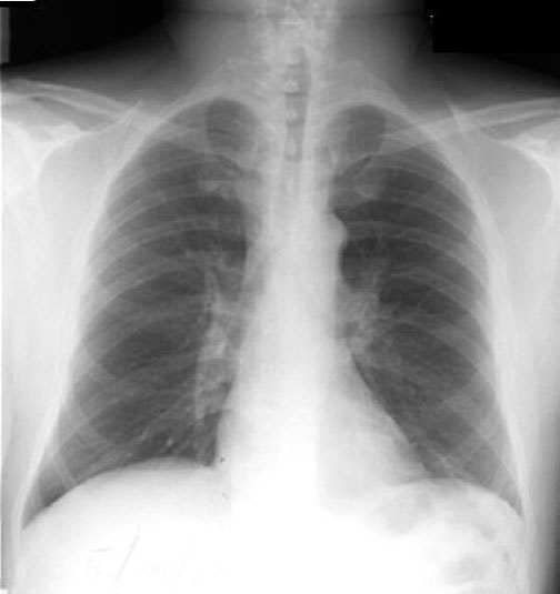
The chest X ray showed a left clavicular hyperostosis and pulmonary cavities

Rapid progression from normal chest x-ray on left. Pneumonia coinciding with

Rapid progression from normal chest x-ray on left. Pneumonia coinciding with

This anteroposterior chest x-ray revealed right upper lobe pneumonia the

This anteroposterior chest x-ray revealed right upper lobe pneumonia the

A chest radiograph was done. After reviewing the findings on that X-ray film

A chest radiograph was done. After reviewing the findings on that X-ray film

This AP chest x-ray shows pneumonia of the left lower lobe with early

This AP chest x-ray shows pneumonia of the left lower lobe with early

 lungs should appear on a chest-x-ray (but not in real life, we hope).
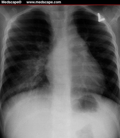
lungs should appear on a chest-x-ray (but not in real life, we hope).

Chest x-ray film of a 6-year-old boy with X-linked recessive chronic
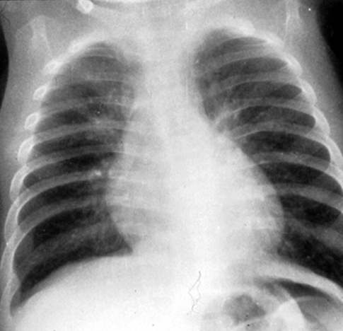
Chest x-ray film of a 6-year-old boy with X-linked recessive chronic

RSV Pneumonia. Chest x-ray 1 There are mild peribronchial infiltrates and

RSV Pneumonia. Chest x-ray 1 There are mild peribronchial infiltrates and

A chest X-ray revealed opacity in the middle third of the right lung field

A chest X-ray revealed opacity in the middle third of the right lung field

This is a chest x-ray of an adult female patient.

This is a chest x-ray of an adult female patient.

Fig 1: Chest x-ray showing collapsed left lung.

Fig 1: Chest x-ray showing collapsed left lung.

Chest x-ray shows diffuse bilateral interstitial opacification.

Chest x-ray shows diffuse bilateral interstitial opacification.

Chest X-ray

Chest X-ray

A chest X ray showing pneumonia in the lower lobe of the patient's right

A chest X ray showing pneumonia in the lower lobe of the patient's right

HX: 4 month old with cough, chest X-ray request says "rule out pneumonia."

HX: 4 month old with cough, chest X-ray request says "rule out pneumonia."

No comments:

Post a Comment